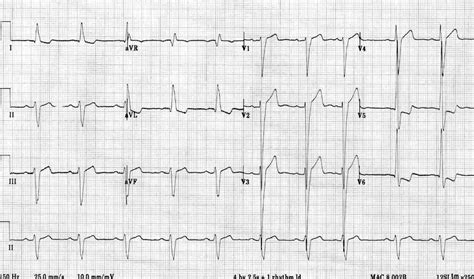
ecg lv hypertrophy | lvh with repolarization abnormality ecg ecg lv hypertrophy The most common causes of left ventricular hypertrophy are aortic stenosis, aortic regurgitation, hypertension, cardiomyopathy and coarctation of the aorta. There are several ECG indexes, which generally have high diagnostic specificity but low sensitivity. LOUIS VUITTON Official USA site - Discover designer women's bags & purses from the world's top luxury Maison. Shop the latest collection of canvas and leather handbags.
0 · what is lvh on ecg
1 · signs of lvh on ecg
2 · lvh with repolarization abnormality ecg
3 · lv hypertrophy ecg criteria
4 · left ventricular hypertrophy with repolarization abnormality
5 · left ventricular hypertrophy life in the fast lane
6 · ecg showing lvh
7 · ecg in left ventricular hypertrophy
For those of you who have requested refunds for Beyond Wonderland SoCal 2020 and have not yet received them, Front Gate will begin processing them by May 7th. For those who have requested a refund for EDC Las Vegas, those refunds will begin processing in about 10 business days. Please bear with us.
The most common causes of left ventricular hypertrophy are aortic stenosis, aortic regurgitation, hypertension, cardiomyopathy and coarctation of the aorta. .

Left ventricular hypertrophy (LVH): Markedly increased LV voltages: huge precordial R and S waves that overlap with the adjacent leads (SV2 + RV6 >> 35 mm). R-wave peak time > 50 ms in V5-6 with associated QRS broadening. LV strain pattern with ST depression and T-wave inversions in I, aVL and V5-6.The most common causes of left ventricular hypertrophy are aortic stenosis, aortic regurgitation, hypertension, cardiomyopathy and coarctation of the aorta. There are several ECG indexes, which generally have high diagnostic specificity but low sensitivity.
Left ventricular hypertrophy changes the structure of the heart and how the heart works. The thickened left ventricle becomes weak and stiff. This prevents the lower left heart chamber from filling properly with blood.
Left ventricular hypertrophy (LVH) refers to an increase in the size of myocardial fibers in the main cardiac pumping chamber. Such hypertrophy is usually the response to a chronic pressure or volume load. The two most common pressure overload states are systemic hypertension and aortic stenosis. Concentric left ventricular hypertrophy is an abnormal increase in left ventricular myocardial mass caused by chronically increased workload on the heart, most commonly resulting from pressure overload-induced by arteriolar vasoconstriction as occurs in, chronic hypertension or aortic stenosis. CONTENTS LAD (left axis deviation) LAHB (left anterior hemiblock) iLBBB (incomplete left bundle branch block) LVH (left ventricular hypertrophy) Diagnostic criteria Interpreting an ECG in the context of LVH LVH versus MI LVH plus ER (early repolarization) Dilated cardiomyopathy HCM (hypertrophic cardiomyopathy) Apical HCM definition of LAD = .
Electrocardiogram. Also called an ECG or EKG, this quick and painless test measures the electrical activity of the heart. During an ECG, sensors called electrodes are attached to the chest and sometimes to the arms or legs. Wires connect the sensors to a machine, which displays or prints results.Current electrocardiographic (ECG) criteria for the diagnosis of left ventricular hypertrophy (LVH) have low sensitivity. Objectives: The goal of this study was to test a new method to improve the diagnostic performance of the electrocardiogram.Left ventricular hypertrophy can be diagnosed on ECG with good specificity. When the myocardium is hypertrophied, there is a larger mass of myocardium for electrical activation to pass.
Left ventricular hypertrophy (LVH) refers to an increase in the size of myocardial fibers in the main cardiac pumping chamber. Such hypertrophy is usually the response to a chronic pressure or volume load. The two most common pressure overload states are . Left ventricular hypertrophy (LVH): Markedly increased LV voltages: huge precordial R and S waves that overlap with the adjacent leads (SV2 + RV6 >> 35 mm). R-wave peak time > 50 ms in V5-6 with associated QRS broadening. LV strain pattern with ST depression and T-wave inversions in I, aVL and V5-6.The most common causes of left ventricular hypertrophy are aortic stenosis, aortic regurgitation, hypertension, cardiomyopathy and coarctation of the aorta. There are several ECG indexes, which generally have high diagnostic specificity but low sensitivity. Left ventricular hypertrophy changes the structure of the heart and how the heart works. The thickened left ventricle becomes weak and stiff. This prevents the lower left heart chamber from filling properly with blood.
Left ventricular hypertrophy (LVH) refers to an increase in the size of myocardial fibers in the main cardiac pumping chamber. Such hypertrophy is usually the response to a chronic pressure or volume load. The two most common pressure overload states are systemic hypertension and aortic stenosis. Concentric left ventricular hypertrophy is an abnormal increase in left ventricular myocardial mass caused by chronically increased workload on the heart, most commonly resulting from pressure overload-induced by arteriolar vasoconstriction as occurs in, chronic hypertension or aortic stenosis.
what is lvh on ecg
CONTENTS LAD (left axis deviation) LAHB (left anterior hemiblock) iLBBB (incomplete left bundle branch block) LVH (left ventricular hypertrophy) Diagnostic criteria Interpreting an ECG in the context of LVH LVH versus MI LVH plus ER (early repolarization) Dilated cardiomyopathy HCM (hypertrophic cardiomyopathy) Apical HCM definition of LAD = . Electrocardiogram. Also called an ECG or EKG, this quick and painless test measures the electrical activity of the heart. During an ECG, sensors called electrodes are attached to the chest and sometimes to the arms or legs. Wires connect the sensors to a machine, which displays or prints results.Current electrocardiographic (ECG) criteria for the diagnosis of left ventricular hypertrophy (LVH) have low sensitivity. Objectives: The goal of this study was to test a new method to improve the diagnostic performance of the electrocardiogram.Left ventricular hypertrophy can be diagnosed on ECG with good specificity. When the myocardium is hypertrophied, there is a larger mass of myocardium for electrical activation to pass.
movado replica watches china
nomos watches replica
signs of lvh on ecg

lvh with repolarization abnormality ecg

Lend Lease – Part 2. Peacekeeping Mission. Learn more. In this guide, we'll talk you through how to unlock Peacekeeper quests in Escape from Tarkov, and run you through all quests he.
ecg lv hypertrophy|lvh with repolarization abnormality ecg


